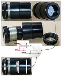
2.1 Ocular micrometer
Introduction
Ocular micrometer is a glass disk that
fits in a microscope eyepiece and that has a ruled scale; when calibrated with
a slide of micrometer and hence the direct measurements of a microscopic object
on prokaryotic and eukaryotic microorganism can be made. It will not change
size when the objectives are changed. Therefore, each objective lens must be
calibrated separately.
Objective
-To measure the cells using a microscope
Materials and Reagents
-Microscope fitted with an ocular micrometer
-Slide micrometer
-Stained preparation of Penicillium
Procedure
1. The
stage micrometer is placed on the stage.
 |
| Figure 2 Stage micrometer |
2. The superimposed image of stage micrometer and eyepiece scale is focused using the objective lens with lowest power which is 4x.
 |
| Figure 3 Microscope |
3. The number of divisions of the eyepiece scale correspond to a definite
number of divisions on the stage scale are determined.
 |
| Figure 4 Ocular scale and stage micrometer scale |
4. The measurement of an eyepiece division in micrometer (µm) is calculated.
5. The experiment is repeated by using the high power.
7. The diameter of the field for each objective was calculated and recorded for future reference.
8. The sample cell’s average dimension in micrometer was determined.
Result
Total magnification = 40x
objective X 10x eyepiece = 400x magnification
 |
| Figure 5 Ocular micrometer and stage micrometer under 400x magnification |
Discussion
1. Ocular micrometer is a glass disc that fit in the
microscope eyepiece that has ruled scale. It is used to measure the size of
magnification object.
2. Stage micrometer is a special glass slide with a known scale.
3. Before we started to measure the size of microorganism, we spent some time to move the stage until the line of ocular micrometer is superimposed to stage micrometer. When the lines of micrometer are coincided, then we are able to measure the size of the microorganism.
4. One division of stage micrometer = 0.01mm.
5. For 400x magnification
Stage scale = 0.01 mm
Let x = 1division of the ocular micrometer
3x = 8 division on stage micrometer x 0.01 mm
3x = 0.08mm
x = 0.0267mm
Therefore, one ocular division = 0.0267mm
Average dimension of sample Penicillium = 0.0267mm x 0.08 = 0.0213mm
Conclusion
Ocular micrometer let us measure
the size of microorganism, Penicillium. Cells can be measured easily
especially using oil immersion.
Reference
http://wiki.answers.com/Q/Why_is_it_necesary_to_calibrate_the_ocular_micrometer_with_each_objective&altQ=Why_it_is_necessary_to_calibrate_ocular_micrometer
http://www.ehow.com/how_5019336_use-ocular-micrometer.html#ixzz1rQeXORkR
http://www.medilexicon.com/medicaldictionary.php?t=55222
http://en.wikipedia.org/wiki/Ocular_micrometer
2.2 Neubauer Chamber
Introduction
To perform counting of cell accurately, Neubauer chamber
is used which is analogous to the quadrat sampling. Neubauer chamber is a thick
glass slide with two counting area separated by a H-shaped depression and it is
0.1mm deep(refer figure 1).A counting special cover slip which is having counting
grid will set on the Neubauer chamber to allow calculation of the concentration
of the cell.
 |
| Figure 1 Neubauer chamber |
Materials and Reagents:
-Neubauer chamber and coverslip
-70% ethanol
-Pipette
Procedure
1. Neubauer chamber was cleaned with 70% ethanol to remove any microorganism or unwanted debris and left it to dry.
2. Neubauer chamber was placed and secured on the stage.
3. The coverslip is placed carefully by 45 degree on the Neubauer chamber.
4. The microscope is focused until an image is formed.
5. The diaphragm is set to smaller to increase image contrast.
Figure
2 The grid layout observed through microscope of the
Discussion
Neubauer chamber consists of finely etched lines that crossing each other perpendicularly which creates a counting grid which aids us in counting the cell. The grid is been seen through 400x of magnification. The grid is divided into 9 large squares. Each square has a surface of area of 1 mm2 and the depth of the chamber is 0.1 mm. Therefore, the volume is 0.9 mm3. There are 16 small squares in a large square. We also have to avoid the present of bubble in the slide of the specimen when we view the grid.
Neubauer chamber consists of finely etched lines that crossing each other perpendicularly which creates a counting grid which aids us in counting the cell. The grid is been seen through 400x of magnification. The grid is divided into 9 large squares. Each square has a surface of area of 1 mm2 and the depth of the chamber is 0.1 mm. Therefore, the volume is 0.9 mm3. There are 16 small squares in a large square. We also have to avoid the present of bubble in the slide of the specimen when we view the grid.
Conclusion
In this experiment, we have learnt to setup the Neubauer
chamber and the method of calculating
the number of cells.


No comments:
Post a Comment